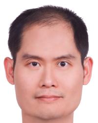
郭柏齡 Po-Ling Kuo
附設醫院復健部兼任主治醫師
Attending Physician in the rehabilitation department at the National Taiwan University Hospital
主要研究領域:
生物物理、力學生物學、生物力學、組織工程﹑醫用超音波Major Research Areas:
Biophysics, Mechanobiology, Biomechanics, Tissue engineering, Medical ultrasound研究領域摘要:
本實驗室主要研究細胞物理學、力學生物學的基礎原理以及相關臨床運用。力學生物學為一新興的跨領域學科,主要探討與力學訊息相關的生物反應。力學訊息目前被認為與多種生理及病理過程有強烈相關,包括組織生成、傷口癒合、血管新生、動脈硬化、心肌肥大、以及腫瘤進展等。因為相對僅能靠擴散方式作用的化學物質而言,力學訊號的作用範圍更遠,傳遞速度也較快。因此在大範圍組織整合過程,包括組織發育、修補、以及退化,惡化,光學訊號可能扮演了具有相當決定性的角色。我們特別對壓力對生物體的影響、生物體如何利用力學訊息通訊、並互相調節功能、以及改造周遭力學環境有興趣。我們研究重點是同質細胞間的自我聚合及功能整合,以及異質細胞間的空間協調。我們的短期目標是發展出能精確測量、並調控細胞與細胞間、以及與介質間力學通訊的實驗平台。遠程目標則是促進吾人對異質細胞間在各種生理、病理狀態下的交互作用,並對組織老化及再生的治療方針上有所啟益。目前本實驗室的研究主題為
- 壓力在細胞生理學以及生物物理學的角色
- 利用生物微機電技術製作可供研究細胞間通訊、以及多重物理因子對細胞生理影響肢體外實驗平台
- 建立可監控細胞與環境力學互動之三維體外實驗平台,並探討該平台在臨床上如藥物篩檢等應用
- 建立臨床上可用於監測及治療緻密結締組織,如肌腱及韌帶,力學功能失常時之非侵入性工具及技術
Research Summary:
Mechanobiology is a new field focusing on understanding how living organisms generate, sense, and respond to various mechanical stimuli, which are believed to play a key role in numerous physiological and pathological processes, such as tissue development, tissue repairing, atherosclerosis, cardiac hypertrophy, and cancer progression. My researches primarily focus on the fundamental mechanisms and clinical applications of mechanobiology. Specifically, we investigate the effects of hydrostatic pressure and environmental elasticity on cell physiology, how cells remodel the mechanical properties of their environment, and develop tools quantitatively evaluate the mechanics of cell-matrix interactions. Our previous achievements and ongoing projects include
1. Elucidate the role of hydrostatic pressure on cell physiology
Hydrostatic pressure is an important physical factor in tissue physiology and pathology. We investigated how hydrostatic pressure affects muscle differentiation, immunological activities, cell motility, and cancer invasiveness. Currently we are working on the possible biological signaling pathways involving these processes.
2. Evaluate the effects of multiple biophysical and biochemical stimuli on cell physiology
The cells in vivo are generally exposed to the coexistence of multiple biophysical and biochemical cues. Knowledge of how cells response to these complex stimuli is important for many disciplines such as regenerative engineering and cancer biology. Using BioMEMS techniques, we have developed several platforms allowing the coexistence of mechanical, electrical, and chemical stimuli for cultured cells. Currently we are delineating the antagonistic and agonistic roles between these stimuli.
3. Develop a 3D cell culture system that allows quantitatively accessing the mechanics of cell-matrix interactions
The changes of mechanical properties such as stiffness of a tissue usually are hallmarks of various physiological and pathological processes, such as arthrosclerosis and tumor malignant transformation. In vitro assays quantitatively measuring the mechanics of cell-matrix interactions are of great importance to understand the mechanisms and facilitate the development of corresponding therapeutic strategies of these processes. Cells cultured in a 3D environment behave far different from that cultured in 2D and recapitulate more physiological characteristics in vivo. An important ongoing project in our lab is to develop a 3D cell culture system using state-of-the-art imaging and scaffold fabrication techniques to quantitatively access the mechanics of live cell-matrix interactions.
4. Develop clinical tools for treatment and monitoring of the mechanical dysfunction of dense connective tissues
Mechanical malfunction of dense fibrous tissues usually leads to protracted and debilitating conditions, such as joint capsule contracture, tissue fibrosis, and tendinosis. Our goal is to develop clinical tools that allow treating these disorders non-invasively, while the change of mechanical function of the diseased tissues can be non-invasively and quantitatively monitored. We have combined the state-of-the-art ultrasonic techniques and developed a prototypical system for this purpose. Our ongoing project is to evaluate its effectiveness in various clinical conditions.

-
B.S.
National Taiwan University, 1994 -
M.S.
National Taiwan University, 1998 -
Ph.D.
Harvard University, 2008
-
Address
MD-519,
Department of Electrical Engineering,
National Taiwan University,
Taipei 106, Taiwan -
Phone
+886-2-33669882 -
Email:

-
Office Hour
Thus 10:00-12:00